
2048R High Speed Linescan Camera
High Speed, High Resolution 2048R SWIR camera with capture rates @ 147 kHz
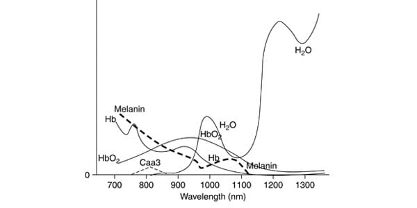
Small animal imaging is important to drug discovery as the effectiveness of drug treatments can be monitored in living animals and studied over time. For instance, photoluminescent taggants can be engineered to latch onto tumor cells, permitting the tumor size to be monitored externally when illuminated by laser light. The laser can be at a shorter NIR wavelength, like 808 nm, but the taggant glow can be at a longer wavelength, such as 1310 nm. The 808 penetrates deeply due to low absorbance, but is the light spreads diffusely due to the scattering. There is still enough excitation energy reaching the taggants to cause emission from longer wavelength. Since the glow is from within the body, the photons only have to make it to the outside, limiting the loss due to water absorbance to just that of one pass through the tissue, while the reduced scattering at longer wavelengths better preserves the shapes of the tagged tumor or organ.
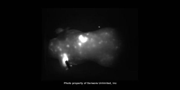
The 1-minute video below illustrates the phenomenon. It was acquired at 60 frames per second by the Sensors Unlimited high-sensitivity SU320HX camera (the most sensitive room temperature InGaAs camera available anywhere). At the start of the video, nanotubes engineered for excitation by the 808 nm laser were injected into the tail of a sedated mouse. The organs light up with 1300 nm light as the nanotubes pass through. The glow periodically increases briefly and one can observe that the animal’s simultaneous physical movement is breathing. This indicates that the intensity flare is possibly due to the increased oxygen from taking the breath. As the movie continues, the vasculature system map of the mouse develops. However, a single image taken after 16 minutes (figure 2) shows this has dissipated and a non-uniform distribution is left, with the concentration of nanotubes saturating the camera in several areas. One of these areas is the ear, seen in the lower left of the image; it has a dark rectangle, showing where a metal identification tag was clipped to the ear.

VIDEO - 58 second movie at 60 fps showing uptake of nanotubes by organs and circulatory system of anesthetized mouse, first appearing at the 3-second point in the midsection, then in the head. Towards the end, the main circulatory vessels are traceable. Note the intensity flares when the mouse takes a breath.
Below is a photoluminescent image 16 minutes after injection of nanotubes. The SU320HX camera sensitivity is unchanged from that of the movie, with the saturated regions indicating very high concentrations have accumulated in just a few places. Use display or editing software to bring up the brightness and reduce the contrast to see the full shape of the mouse. The dark rectangle on the bright area in the lower left is a metal identification tag clipped to the mouse’s left ear.